Die Entdeckung von Biomarkern kann zu einem Bluttest für Hirntumoren führen
[ad_1]
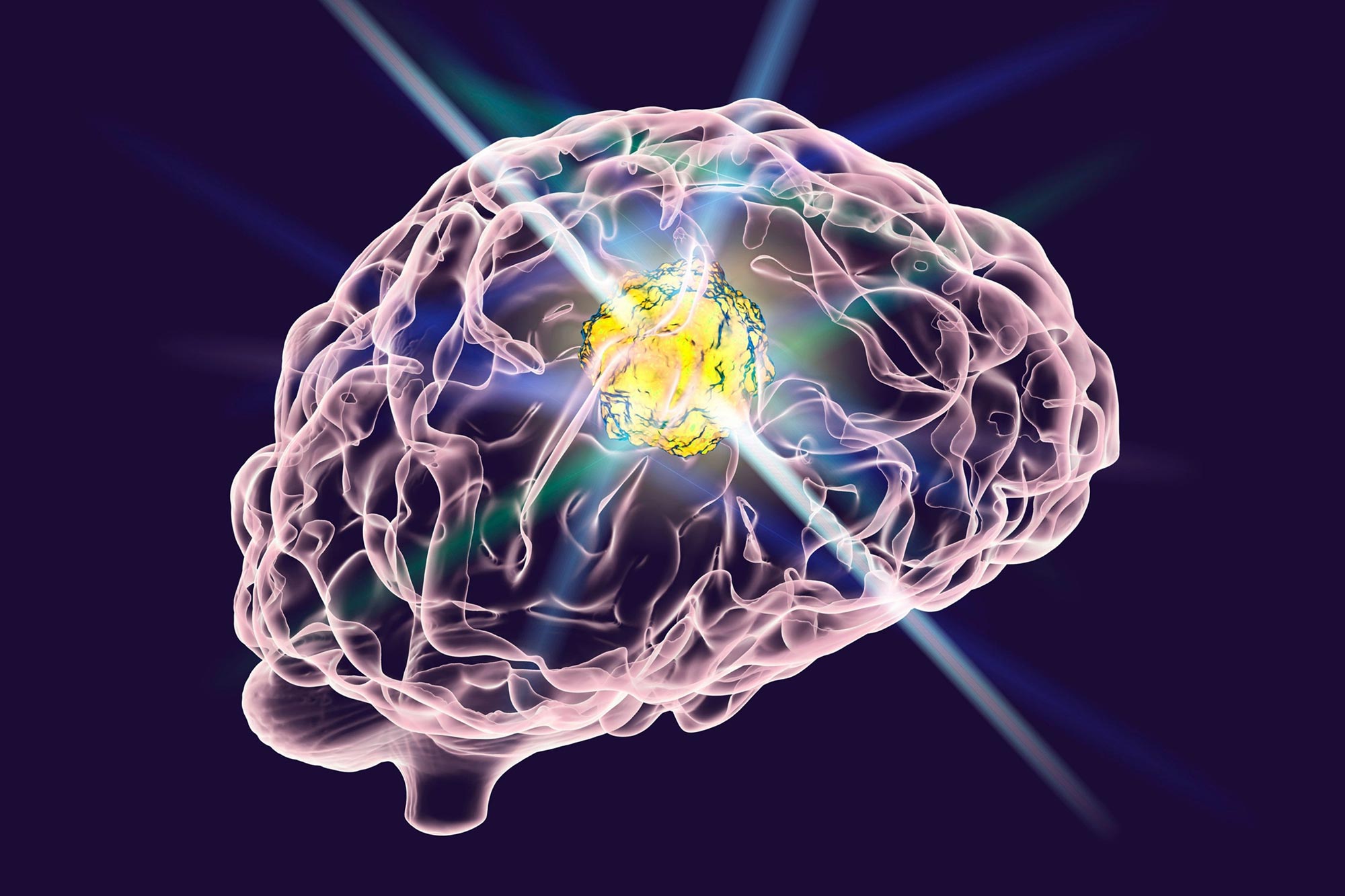
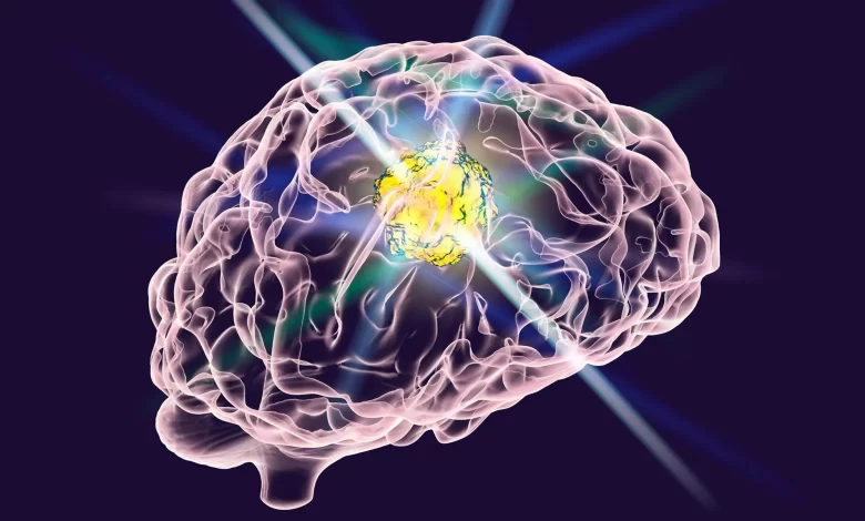
Forscher des Penn State College of Medicine haben einen Biomarker identifiziert, der in Bluttests zur Diagnose des Glioblastoms, der häufigsten und tödlichsten Art von Hirntumoren, verwendet werden kann, und um sein Fortschreiten zu verfolgen und die Behandlung zu steuern.
Das Glioblastom (GBM) ist die häufigste und tödlichste Form von Hirntumoren mit einer Fünf-Jahres-Überlebensrate von nur 5 %. Forscher des Penn State College of Medicine haben einen Biomarker identifiziert, der in Bluttests verwendet werden kann, um GBM zu diagnostizieren, seinen Fortschritt zu verfolgen und die Behandlung zu steuern. Die Forscher sagten, dass eine solche nicht-invasive Flüssigbiopsie für GBM Patienten helfen könnte, schneller die Versorgung zu erhalten, die sie benötigen.
„Patienten erhalten normalerweise bildgebende Verfahren wie MRT- oder CT-Scans, um das Fortschreiten von Hirntumoren zu diagnostizieren und zu verfolgen, aber es kann für Ärzte schwierig sein, anhand dieser Scans zu erkennen, ob es dem Patienten besser oder schlechter geht, da sie keine Details liefern auf zellulärer oder molekularer Ebene“, sagte Vladimir Khristov, Absolvent und Medizinstudent, Penn State. „Deshalb brauchen wir einen ergänzenden diagnostischen Test, der Ärzten hilft, festzustellen, ob die Tumore auf die Therapie ansprechen und sich zurückbilden, oder ob sie sich verschlimmern und eine zusätzliche Behandlung benötigen.“
In der Tat, fügte Brad Zacharia, außerordentlicher Professor für Neurochirurgie und HNO-Heilkunde, Penn State, hinzu, könnte eine Flüssigbiopsie für Glioblastome von enormem Wert für Patienten sein, die an diesem verheerenden Tumor leiden.
„Eine Flüssigbiopsie kann die Diagnose erleichtern und, was noch wichtiger ist, ein besseres Verständnis der Reaktion des Tumors auf die Behandlung auf eine Weise liefern, die unseren derzeitigen Technologien fehlt“, sagte er.
Das Team untersuchte einen bestimmten Antigenrezeptor namens Interleukin-13-Rezeptor α2 (IL13Rα2), von dem bekannt ist, dass er im Tumorgewebe von mehr als 75 % der GBM-Patienten erhöht ist.
„Obwohl es in Tumorgewebe signifikant überexprimiert wird, haben keine Studien das diagnostische und prognostische Potenzial von IL13Rα2 untersucht, das in Bioflüssigkeiten von Patienten zirkuliert“, sagte James Connor, angesehener Professor für Neurowissenschaften und Anatomie, Penn State.
Um die Nützlichkeit von IL13Rα2 als Biomarker für GBM zu untersuchen, untersuchten die Forscher das Tumorgewebe und Blut[{” attribute=””>plasma of 79 patients with primary GBM, along with the blood plasma of 23 control patients, from two different health systems. The control patients had primary diagnoses of either spinal stenosis or arteriovenous malformation but did not have any malignancy or chronic inflammation.
In the patients’ plasma, the researchers looked specifically at extracellular vesicles, which are small particles that are released by cells and carry material from those cells. They found that patients with GBM had significantly elevated levels of IL13Rα2 in their blood plasma compared to control patients and that the IL13Rα2 was likely concentrated on extracellular vesicles derived from tumor cells. They also found that these IL13Rα2 levels in blood plasma were correlated with the IL13Rα2 levels in the patients’ tumors. Their findings were published recently in the Journal of Neuro-Oncology.
“The fact that we documented IL13Rα2 on tumor-derived extracellular vesicles and that we observed a correlation between plasma and tumor levels of IL13Rα2 suggests that plasma IL13Rα2 does indeed derive from GBM tumors,” said Khristov. “This is important because previously it was difficult to tell if the IL13Rα2 in plasma came from the tumors, or if they came from the body’s response to the tumors. Our findings suggest that IL13Rα2 does have utility as a biomarker for glioblastoma.”
Connor noted that the finding is especially significant given that IL13Rα2 has been shown to have a patchy distribution in GBM tumors, raising the question of whether a needle biopsy or small sample of tumor tissue is representative of the tumor as a whole.
“Testing for IL13Rα2 circulating in plasma may provide an even better picture of the presence and extent of GBM than a tumor sample,” said Connor. Additionally, he said, “the tumor-specific nature of IL13Rα2 implies that it can be used for tumor-targeted therapies without affecting outside tissues.”
Interestingly, the team found that elevated levels of IL13Rα2 in both plasma and tumors predicted longer overall survival. In fact, patients with high levels of plasma IL13Rα2 had a 6.5-month longer median overall survival compared to patients with low levels.
“It seems counterintuitive that high levels of plasma IL13Rα would confer a survival advantage since their presence indicates a tumor and, ultimately, we do not know why this is the case,” said Khristov. “However, there is some evidence that increased IL13Rα2 is correlated increased fibrosis in the tumor, which indicates tissue healing. It’s important for patients to know if they may have this survival advantage or not.”
Zacharia noted that this work, and that of many other studies, relies on biological specimens, such as blood, tumor tissue and spinal fluid, from patients.
“Their generous and selfless gifts of these specimens to the Penn State Neuroscience Institute Biorepository make this work possible,” he said, “and we are forever grateful to the patients and their families.”
Reference: “Plasma IL13Rα2 as a novel liquid biopsy biomarker for glioblastoma” by Vladimir Khristov, Darya Nesterova, Mara Trifoi, Taylor Clegg, Annika Daya, Thomas Barrett, Emily Tufano, Ganesh Shenoy, Bhavyata Pandya, Gela Beselia, Nataliya Smith, Oliver Mrowczynski, Brad Zacharia, Kristin Waite, Justin Lathia, Jill Barnholtz-Sloan and James Connor, 27 November 2022, Journal of Neuro-Oncology.
DOI: 10.1007/s11060-022-04196-0
Other Penn State authors on the paper include Darya Nesterova, general surgery resident; Mara Trifoi, medical student; Taylor Clegg, medical student; Annika Daya, medical student; Thomas Barrett, medical student; Emily Tufano, graduate student; Ganesh Shenoy, medical student and Ph.D. student, Bhavyata Pandya, graduate student; Nataliya Smith, human research technologist; Oliver Mrowczynski, neurosurgery resident; Brad Zacharia, associate professor of neurosurgery and otolaryngology; Gela Beselia, postdoctoral fellow, Albany Medical College; Kristin Waite, staff scientist, National Cancer Institute; Justin Lathia, professor of molecular medicine, Case Western Reserve University; Jill Barnholtz-Sloan, senior investigator, National Cancer Institute.
[ad_2]
Source link